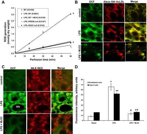
Fig. 1.
Reactive oxygen species (ROS) production in isolated perfused mouse lungs and lung cells in situ after LPS ± 1-hexadecyl-3-(trifluoroethyl)-sn-glycero-2-phosphomethanol (MJ33). LPS at 1 mg/kg and liposomes containing 4 nmol MJ33 were concurrently administered intratracheally (IT) to the intact mouse. A: ROS generation by isolated perfused wild-type (WT), NADPH oxidase type 2 (NOX2)-null, and peroxiredoxin 6 (Prdx6)-null mouse lungs was measured by Amplex Red oxidation. Perfusate samples were taken at intervals and analyzed for oxidized Amplex Red; results are plotted vs. time of perfusion. A perfusate sample from time zero was used as the baseline. The numbers in parentheses indicate the slope of the line (nmol/g dry wt per min) calculated by the least mean squares method. Values represent mean ± SE for n = 3; in some cases the SE bars are within plotted points. *P < 0.01 compared with WT at 60 min perfusion time. Effect of MJ33 on LPS-induced ROS in endothelial cells (B) and type 2 epithelial cells (C) of mouse lungs by confocal microscopy. Fluorescent dyes were used to detect ROS (dichloroflorescein, DCF; green color) and to identify cells (red color), either endothelial cells (Alexa 594AcLDL) or epithelial cells (Nile Red). The yellow areas indicate colocalization of DCF fluorescence with the cell marker. The dark areas are the alveolar spaces (Alv). D: quantification of the effects of MJ33 on LPS-induced ROS in endothelial cells and lung alveolar type II cells from the isolated perfused mouse lungs. Data represent the means ± SE for n = 3 independent experiments. Each experiment utilized a separate perfused lung, and for each 3 randomly selected fields were imaged, the integrated fluorescence was determined, and the values were averaged. Studies evaluated 3 treatment groups (basal, LPS, and LPS + MJ33). *P < 0.05, **P < 0.01 compared with basal and #P < 0.05, ##P < 0.01 compared with LPS.

