Fig. 3.
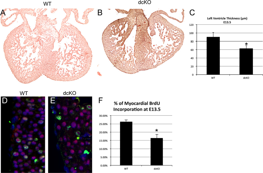
dcKO hearts display myocardial thinning and decreased proliferation. Hematoxylin and eosin staining was used to visualize the morphology of wild type (A) and dcKO (B) hearts at E13.5. We observed myocardial thinning in dcKO hearts (B) when compared to wild type hearts (A). Measurement of left ventricle thickness revealed a significant decrease between dcKO hearts that displayed thinning and wild type hearts (* = p < 0.05, (C)). To measure myocardial proliferation, BrdU incorporation (Green) was quantified in Nkx2.5 (Red) expressing cardiomyocytes (D)–(E).We found a statistically significant decrease (* = p < 0.005) in the percentage of BrdU positive cells within the myocardium of dcKO hearts when compared to wild type hearts (F). Images were counterstained with DAPI (Blue) to label nuclei. Error bars represent standard deviation. (For interpretation of the references to color in this figure legend, the reader is referred to the web version of this article.)

