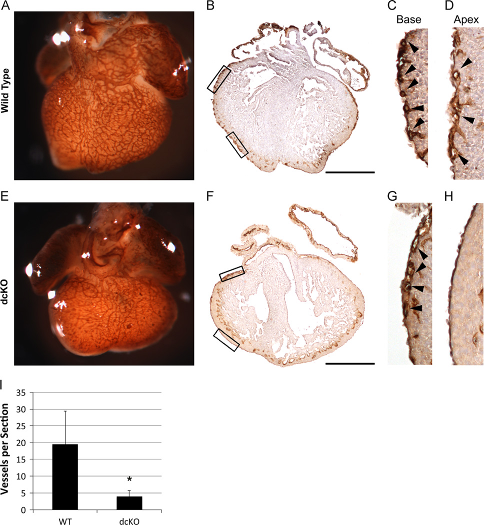
Fig. 4.
dcKO hearts exhibit decreased coronary plexus formation. Whole mount PECAM staining was used to visualize the formation of the coronary plexus at E13.5 (A) and (E). dcKO hearts (E) display decreased coverage of the ventricle when compared to wild type hearts (A). Sections prepared from whole mount PECAM stained hearts were used to visualize the formation of the coronary plexus at E13.5 (B)–(D), (F)–(H). Wild type hearts (B) display extensive sub-epicardial vessel formation, while dcKO hearts (F) show very few vessels. Higher magnification of outlined boxes in whole section images show vessels at the base of both wild type (C) and dcKO hearts (G); however, the spreading of vessels towards the apex of the wild type heart (D) is not replicated in the dcKO (H). Black arrowheads (C), (D), (G) and (H) mark sub-epicardial vessels. Decreased PECAM signal in endocardium (B)–(D) and (F)–(G) is due to limited penetrance of whole mount staining. (I) Quantification of vessels per section revealed a statistically significant (* = p < 0.05) decrease in dcKO hearts when compared to wild type hearts. Error bars represent standard deviation. Scale bars represent 500 µm.

