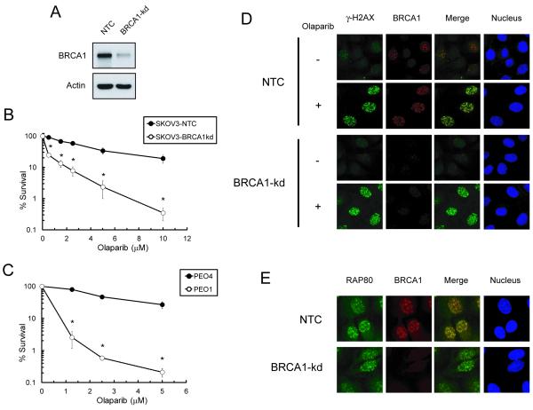
Fig. 1. Lack of BRCA1 foci formation and enhancement of olaparib sensitivity in BRCA deficient EOC cell lines.
A. Western blot analysis of BRCA1 levels in non-targeted siRNA control (NTC) and BRCA1-knockdown (BRCA1-kd) SKOV-3 cells. B. SKOV3 cells and C. PEO1 and PEO4 cells were exposed continuously to various concentrations of olaparib and clonogenic survival was determined. Data are means ± SD. * p<0.05 compared to NTC SKOV-3 cells at each concentration. D. Cells were untreated or treated with 5 μM olaparib for 6 hr. Immunofluorescence of γ-H2AX (green), BRCA1 (red) foci, and nuclei (blue) was visualized by confocal microscopy. E. Cells treated with 5 μM olaparib for 6 hr are shown for immunofluorescence of RAP80 (green), BRCA1 (red) foci, and nucleus (blue).

