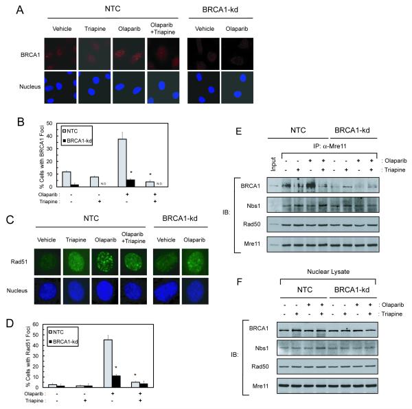
Fig. 3. Triapine abrogates olaparib-induced nuclear repair foci and disrupts BRCA1 interaction with the MRN complex in BRCA1 wild-type SKOV-3 cells.
A, B. NTC and BRCA1-kd SKOV-3 cells were treated with 5 μM olaparib, 0.75 μM triapine, or both agents in combination for 6 hr. Cells were stained with an anti-BRCA1 antibody and nuclei were counterstained. Immunofluorescence of BRCA1 foci (red) and nuclei (blue) were visualized by confocal microscopy. Cells were scored for nuclei containing more than ten BRCA1 foci to determine the % cells with positive BRCA1 foci from 5-10 random fields. N.D. indicates no detectable foci. Data are means ± SD. * p<0.05 compared to NTC cells treated with olaparib. C, D. Cells were treated as described in A. Cells were stained with an anti-Rad51 antibody and nuclei were counterstained. Immunofluorescence of Rad51 foci (green) and nuclei (blue) were visualized by confocal microscopy. Immunofluorescence of Rad51 foci in a single cell is shown. Cells were also scored for nuclei containing more than ten distinct Rad51 foci to determine the % cells positive for Rad51 foci. Data are means ± SD. * p<0.05 compared to NTC cells treated with olaparib. E. NTC and BRCA1-kd SKOV-3 cells were treated with 5 μM olaparib, 0.75 μM triapine, or both agents in combination for 6 hr. Nuclear protein was isolated for co-immunoprecipitation with an anti-Mre11 antibody. Immunoprecipitates were analyzed for the protein levels of BRCA1, Nbs1, Rad50, and Mre11 by western blotting. Ten μg of nuclear protein from untreated NTC cells were also included as an input for positive controls in the analysis. F. Nuclear protein was analyzed by western blotting using BRCA1, Nbs1, Rad50, and Mre11 antibodies to demonstrate approximately equal amounts of protein in all lysate samples used for co-immunoprecipitation.

