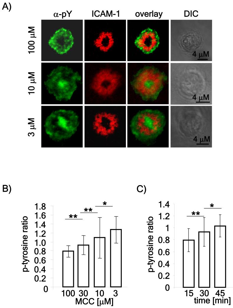
Figure 1. Detection of tyrosine phosphorylation in the cSMAC using low doses of peptide.
A) Naïve CD4 T cells were isolated from spleens of AND mice, stimulated with MCC loaded, irradiated B10.BR splenocytes for 5 days and used on day 6. The rested AND T cells were flowed over lipid bilayers loaded with the indicated doses of MCC for one hour after which the bilayers were fixed, permeabilized and stained using an antibody to phosphotyrosine. The images are representative of over 50 cells obtained in three independent experiments. B) Phosphotyrosine staining in the cSMAC and pSMAC of more than 50 cells obtained in three independent experiments was measured using ImageJ software and represented as the ratio between phosphotyrosine staining in the cSMAC compared to the pSMAC. C) T cells were allowed to form mature synapses for 15, 30 or 45 minutes on lipid bilayers loaded with 3 μM MCC. The bilayers were stained and quantitated as described above. P values (* <0.05; ** < 0.01) were obtained using the Student’s t test.

