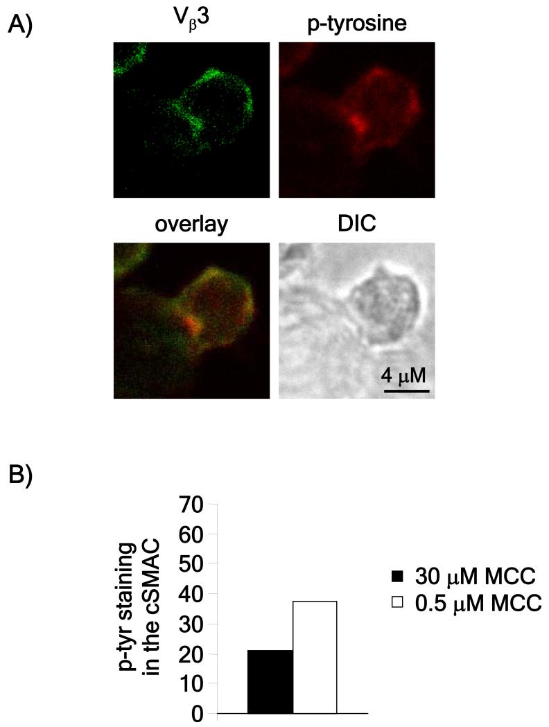
Figure 2. Detection of tyrosine phosphorylation in T cell-APC conjugates.
A) Rested AND T cells were stimulated with CH27 cells loaded with the indicated amounts of peptide for one hour. Conjugates were then fixed, permeabilized and stained using antibodies to Vβ3 and phosphotyrosine. The images are representative of over 20 conjugates obtained in two independent experiments. B) Phosphotyrosine staining in the cSMAC was measured using ImageJ software and represented as the percentage of cells in which phospho-tyrosine staining in the cSMAC was at least 1.5 fold higher compared to the pSMAC (the area of the contact site excluding the cSMAC). Conjugates in which Vβ3 accumulation in the central third of the contact site was at least 1.5 times higher than at the rest of the contact site were considered to have cSMACs.

