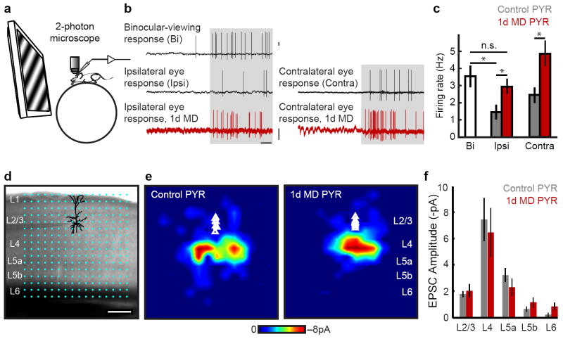
Figure 1. L2/3 pyramidal neuron responsiveness and local circuit organization is unchanged 1d after MD.
a-c, Responses of pyramidal (PYR) neurons to drifting gratings in alert mice. a, Cartoon of head-fixed configuration. b, Example loose-cell attached recordings from controls (black) and after 1d MD (red) in response to visual stimulation (gray shading). Scale: 1mV, 500 ms. c, Mean firing rate at optimal orientation (Bi 10 mice, n= 30 cells; Ipsi control 7 mice, n= 22 cells; Ipsi MD 6 mice, n= 33 cells; Contra control 3 mice, n= 9 cells; Contra MD 3 mice, n= 10 cells). d, PYR neuron recorded in binocular V1 in an acute slice; overlaid are 16 ×16 LSPS stimulation locations spanning pia to white matter. e, In vitro LSPS aggregate excitatory input maps pooled across PYR neurons, triangles indicate soma location (Control 4 mice, n= 9 cells; MD 4 mice, n= 9 cells). Scale: 200 μm. f, Mean LSPS-evoked EPSC amplitude, same cells as e. *P<0.05.

