Figure 5.

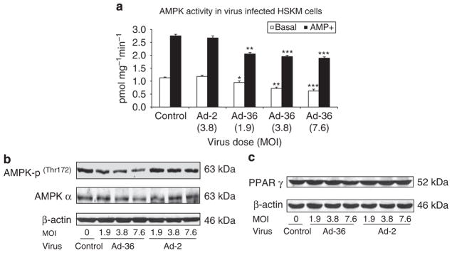
Ad-36 affects AMPK signaling in muscle cells. At day 6 after virus infection, cells were harvested and 200 μg cell lysates were immunoprecipitated with antibody against AMPK α and protein A. AMPK activity was carried out by adding 200 μM of [γ32P]ATP and SAMS peptide with or without 200 μg AMP and incubated at 32 °C for 10 min. Reaction was terminated by spotting a 20 μl aliquot onto a 1 cm2 piece of P-81 phosphocellulose paper and washing for 3×5min in 1% phosphoric acid. Samples were then air-dried, incorporation of γ-32P was quantified and AMPK activity is expressed as incorporated ATP (pmols) per mg protein per min. (a) AMPK activity in virus-infected and control muscle cells. Assay was conducted in triplicate. Mean±s.e.m., *P<0.05, **P<0.01 and ***P<0.001, Ad-36 vs control. (b) AMPK-p and AMPK α protein as well as PPAR γ (c) abundance was measured by western blot in virus-infected and non-infected control muscle cells.
