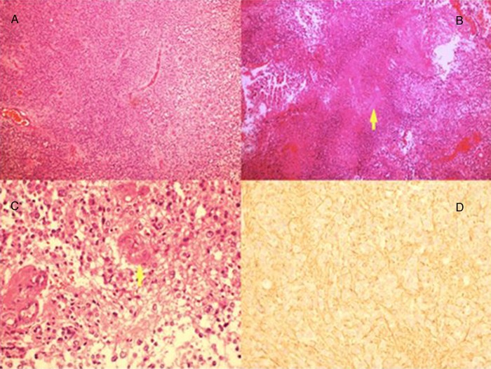
Figure 2.
(A) H&E-stained section at ×10 magnification showing nests of tumour cells arranged around blood vessels forming rosettes. Individual cells are small with rounded hyperchromatic nuclei. (B) Tumour cells with extensive necrotic area. (C) H&E-stained section showing tumour cells with nuclear atypia and pleomorphism. There is a focus of endothelial hyperplasia in a small vessel. (D) Glial fibrillary acidic protein immunohistochemical stain which is negative in tumour cells but positive in fibrillary processes around them.

