
Figure 1. Brain MRI.
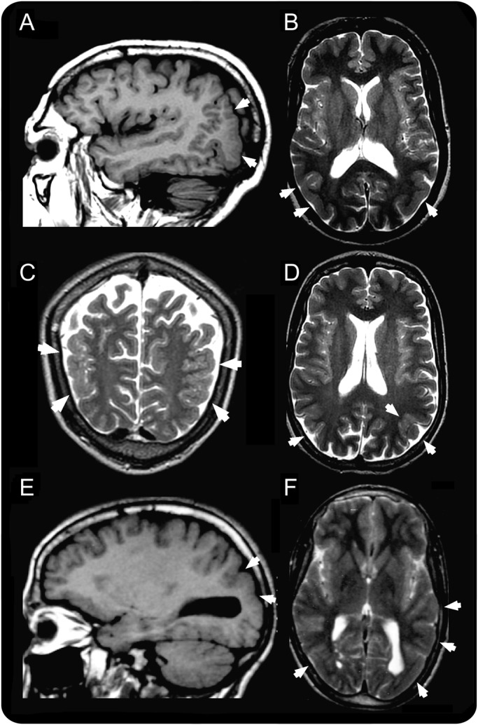
(A, B) Patient V.8, (C, D) patient V.9, and (E, F) patient V.11. Images A and E are T1-weighted sagittal sections; B, D, and F are T2-weighted axial sections; and C is a T2-weighted coronal section. The areas of abnormal cortical development in the temporo-occipital areas are indicated by the white arrows and are consistent in all 3 patients with a combination of increased cortical thickness, smooth cortex, overfolding, and microgyri.