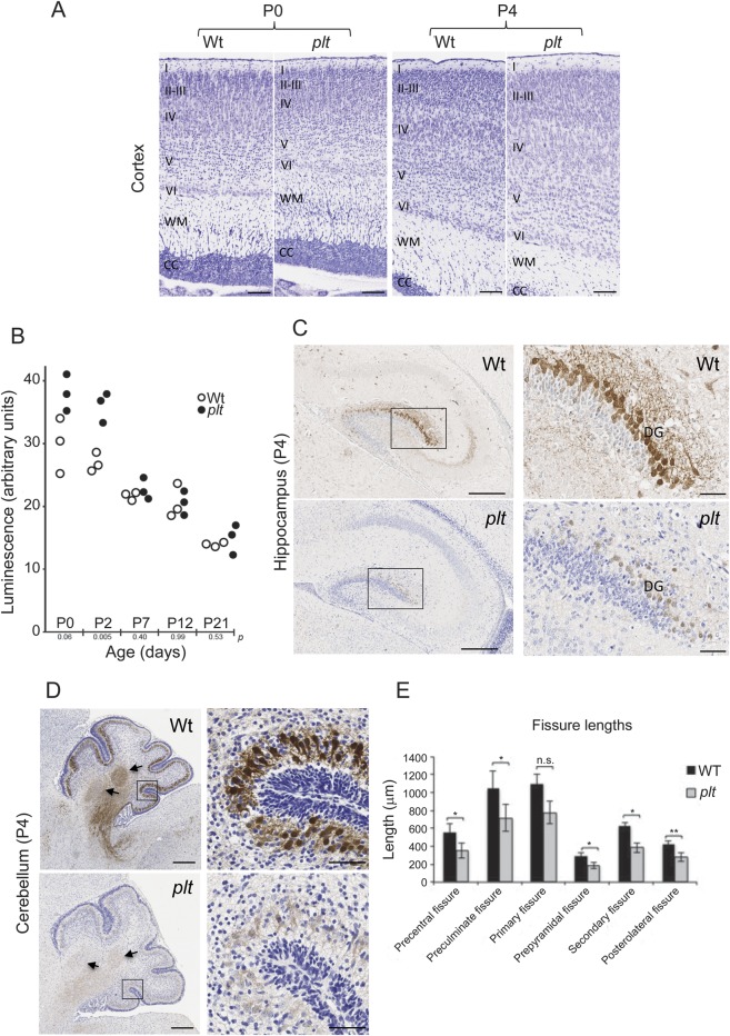
Figure 3. Neurodevelopmental anomalies in plt mice.
(A) Nissl staining of cortex from plt homozygote and WT littermate at postnatal days P0 and P4. Note presence of swollen neurons in layers IV and V of plt aged P4. Cortical layers are indicated as follows: I–VI, WM (white matter), CC (corpus callosum). (B) Caspase 3/7 activity was measured in brain homogenates from 3 animals of each genotype at the indicated postnatal age. An unpaired Student t test was used to generate p values at each age. (C) Weak calbindin immunoreactivity in hippocampus of plt mouse compared with WT littermates. Higher magnification of dorsal DG (boxed regions) shows many granule cells with a well-developed dendrite tree in the WT brain, while in the plt, there is weak staining of the perikaryon and no immunostaining of dendrite trees. (D) Very weak calbindin immunoreactivity in cerebellar cortex and deep nucleus (arrows) in plt mouse compared with WT littermate at P4. Boxed areas are focused on posterolateral fissures. Scale bars: A, 100 μm; C, 300 μm (boxed area 50 μm); D, 250 μm (boxed area 50 μm). (E) Histogram representing the fissure lengths in the cerebellum at P4 (n = 5). Significant differences between lengths of fissures were observed between WT and plt. *p < 0.05; **p < 0.01. DG = dentate gyrus; n.s. = not significant; plt = pale tremor; WT = wild-type.

