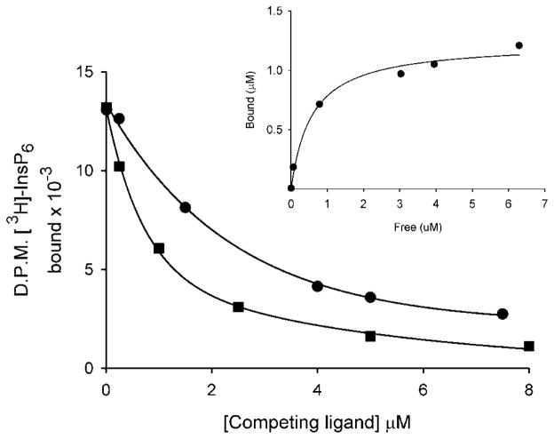
Figure 3. Use of a PEG precipitation assay to identify binding of InsP6 and PtdIns(3,4,5)P3 to PBD2.
PBD2 (32 μg) was incubated with 3HInsP6 and increasing amounts of either non-radioactive InsP6 (circles) or C8 -PtdIns(3,4,5)P3 (squares) as described in the Materials and methods section. Bound and free ligand was separated by a PEG precipitation technique (see Materials and methods section). The Kd for InsP6 (0.57 μM) was calculated from the saturation binding plot (inset). The value of Bmax was 0.6 mol/mol of protein.

