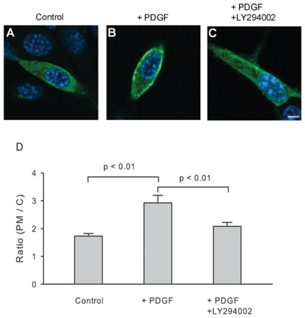
Figure 8. PDGF promotes translocation of PPIP5K to the plasma membrane in NIH 3T3 cells.
At 1 day after transfecting NIH 3T3 cells with cDNA encoding FLAG-tagged PPIP5K1, the intracellular location of the enzyme was determined by confocal immunofluorescence microscopy. (A) Control. (B) Treatment with 50 ng/ml PDGF for 5 min. (C) 15 min pre-treatment with 100 μM LY294002 [50] before 50 ng/ml PDGF was added for 5 min. The scale bar represents 5 μm. (D) The concentration of PPIP5K at the plasma membrane (PM) is shown as a ratio to the concentration of kinase in the cytosol (C) [30]. Results are the means ± S.E.M. from 31–34 cells.

