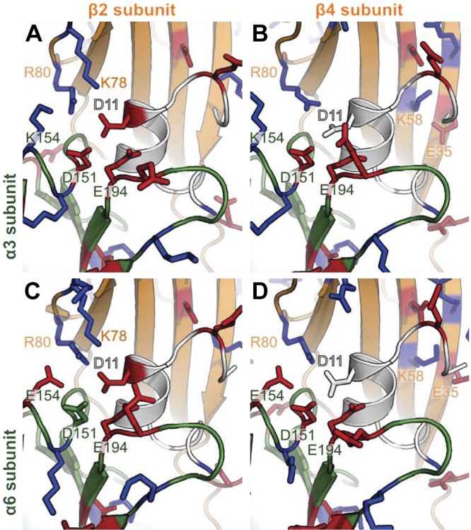
Figure 8.

Homology models of the interactions involving α-CTx LvIA and α3β2 nAChR (A), α3β4 nAChR (B), α6β2β3 nAChR (C), and α6β4 nAChR (D). LvIA is in white, α3 and α6 subunits are in dark green, and β2 and β4 subunits are in orange. Positively and negatively charged residues, as predicted by PROPKA3.1 at pH 7.0, are shown as blue and red sticks, respectively. Important residues that were not predicted to be charged in some complexes, including D11 and D151, are shown as sticks. Numbering of positions is according to the α3 and β2 ligand-binding domains.
