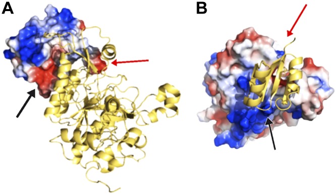
Figure 4.

Complementary electrostatic charge interactions of the surfaces of the prodomain and protease domain in rEpiP protein. A) Prodomain as an electrostatic surface and the protease domain as a ribbon diagram. B) Prodomain as ribbon diagram and protease domain is an electrostatic surface. Arrows indicate corresponding interactions of the 2 domains in each panel. Electrostatic surfaces are shown in red for negative charge, blue for positive charge, and white for neutral charge.
