Figure 4.
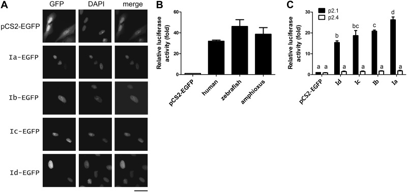
All 4 amphioxus HIFα isoforms were nuclear proteins capable of activating HRE-dependent gene expression. A) Nuclear localization. U2OS cells were transfected with the indicated pCS2 plasmids. Left panels: EGFP signal was visualized 48 h after transfection. Middle panels: corresponding DAPI staining. Right panels: merged images. Scale bar = 50 μm. B) Amphioxus HIFα Ia had transactivation activity comparable to those of human and zebrafish HIF-1α. C) Transactivation activities of amphioxus HIFα isoforms. HEK 293T cells were transfected with pCS2-EGFP or the indicated HIFα expression vectors together with the p2.1 reporter (solid bars) or the p2.4 (open bars) plasmids. pRL-SV40T (Renilla) was cotransfected as an internal control. Luciferase activity was measured at 24 h after the transfection. Transactivation activity is expressed as fold increase over the pCS2-EGFP control group. Values are means ± sd (n=3). Groups with different letters differ significantly from each other (P<0.05).

