
Figure 7.
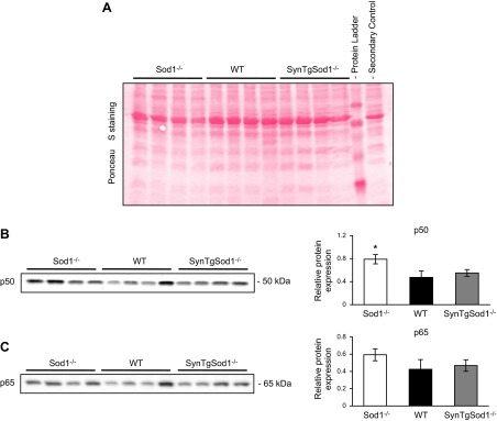
NFκB activation assessed by nuclear location of the p50 and p65 subunits of NFκB in skeletal muscle of SynTgSod1−/− mice. A) Ponceau S staining used to ensure equivalent loading of the samples, corresponding to the Western blots in panels B, C. B, C) Representative Western blots of the NFκB p50 (B) and p65 (C) subunits in nuclear fractions from skeletal muscle of Sod1−/−, WT, and SynTgSod1−/− mice and densitometric quantification of the blots. *P < 0.05 vs. WT.