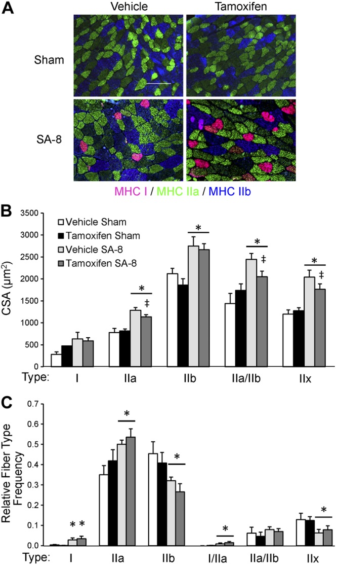
Figure 3.

Plantaris fiber type shift 8 wk following SA surgery (SA-8) is independent of satellite cell content. A) Immunohistochemical analysis of plantaris muscle cross sections for myosin heavy chain (MHC) type I (pink), type IIa (green) and type IIb (blue). Unstained fibers are MHC type IIx. Scale bar = 100 μm. B) Quantification of MHC-specific fiber CSA, presented as mean ± se CSA (μm2); n = 6. C) Quantification of the relative frequency of the different fiber types, presented as mean ± se relative frequency; n = 6. *P < 0.05 for surgery between condition-matched groups; ‡P = 0.1 for tamoxifen between condition-matched groups (trend toward significant effect).
