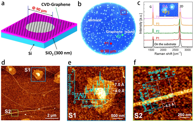
Figure 1. Preparation and characterizations of the suspended CVD-graphene membrane.
(a), Schematic of the freely suspended CVD-graphene on SiO2 substrate with a cylindrical hole. (b), Optical microscope image of the suspended monolayer graphene with wrinkles and graphene islands. (c), Comparison of Raman spectra (excitation wavelength λ = 514 nm) measured on various locations, P1: monolayer graphene, P2: wrinkle, P3 and P4: graphene island. (d–f), Contact mode AFM images of the graphene with wrinkles and islands, which were measured at the supported graphene on Si substrate near the suspended graphene.

