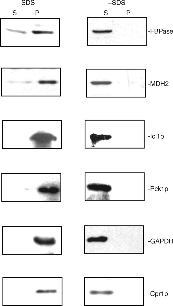
Fig. 2.
FBPase, MDH2, Icl1p, Pck1p, GAPDH, and Cpr1p are distributed in the vesicle-enriched fraction. Wild-type cells were starved of glucose for 3 days and harvested. Total extracts were obtained and centrifuged at 3,000×g for 5 min. The resulting supernatant was further centrifuged at 200,000×g for 2 hours. The 200,000×g pellet fraction was resuspended, aliquoted, and incubated in the absence or presence of 2% SDS for 30 min. Following incubation, samples were re-centrifuged at 200,000×g for 2 hours. The distribution of FBPase, MDH2, Icl1p, Pck1p, GAPDH, and Cpr1p in the 200,000×g supernatant (S) and pellet (P) fractions were examined by Western blotting. Representative data from 3 experiments are shown.

