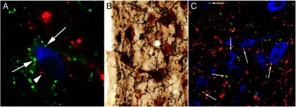
Figure 5.

Innervation of the TRH neurons in the rat PVN by axons originating from the arcuate nucleus (A), DMN (B), and catecholaminergic neurons (C) in the brainstem. A, TRH neurons (blue) are contacted by axon terminals containing α-MSH (red; arrowhead) and AGRP (green; arrows). B, Axon varicosities containing the anterogradely transported marker protein, PHA-L (black) are juxtaposed to TRH-synthesizing neurons (brown) after iontophoretic administration of the tracer into the DMN. C, Both noradrenergic (red; open arrows) and adrenergic (yellow, white arrows) axons establish contacts with the TRH neurons. [Modified from C. Fekete et al: α-Melanocyte-stimulating hormone is contained in nerve terminals innervating thyrotropin-releasing hormone-synthesizing neurons in the hypothalamic paraventricular nucleus and prevents fasting-induced suppression of prothyrotropin-releasing hormone gene expression. J Neurosci. 2000;20:1550–1558 (52), with permission. © Society for Neuroscience. From E. Mihály et al: Hypothalamic dorsomedial nucleus neurons innervate thyrotropin-releasing hormone-synthesizing neurons in the paraventricular nucleus. Brain Res. 2001;891:20–31 (74), with permission. © Elsevier. And from T. Füzesi et al: Noradrenergic innervation of hypophysiotropic thyrotropin-releasing hormone-synthesizing neurons in rats. Brain Res. 2009;1294:38–44 (61), with permission. © Elsevier.]
