
Figure 4.
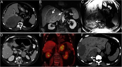
A, CT of ACC showing large heterogeneous right adrenal tumor, B, Contrast-enhanced coronal MRI in the same patient showing heterogeneous enhancement with nonenhancing areas of necrosis (arrows). C, Non–contrast-enhanced T1-weighted MRI in the same patient showing T1-weighted hyperintense areas of hemorrhage (arrows). D, CT of ACC showing left adrenal tumor. E, Intensely FDG avid left adrenal mass in the same patient. F, Metastasized ACC, calcifications in primary tumor (arrows).