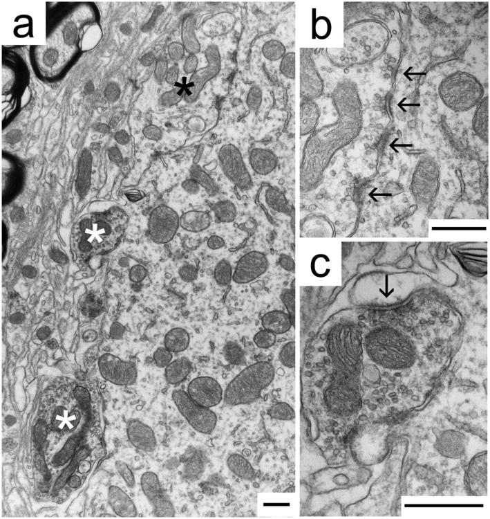
Figure 4.
Immunohistochemical labeling of synaptic endings using GlyT2 antibodies identifies inhibitory synaptic contacts (white asterisks, c) on a representative MSO cell body of a cat with normal hearing (a). The immunoprecipitate darkens the mitochondria and forms a thin, dark halo around SVs, clearly revealing unstained terminals (black asterisk, b). b: A typical GlyT2-immunonegative ending has round SVs and asymmetric postsynaptic densities (arrows) characteristic of excitatory synapses. c: A typical GlyT2-immunopositive terminal has pleomorphic SVs and symmetric postsynaptic densities (arrow), characteristic of inhibitory synapses. Scale bars = 500 nm.

