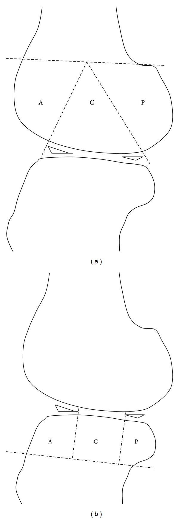
Figure 1.

A sketch of the subregions over the femoral (a) and tibial (b) condyles tailored for the measurement of cartilage defect on sagittal histomorphological sections. Both of the femoral and tibial articular surfaces were divided into the anterior (A), central (C), and posterior (P) subregions. Region A of the femur corresponded to the patellofemoral articulation; region C represented the weight-bearing surface, and region P represented the posterior convexity, which articulates only in extreme flexion. Region C of the tibial surface represented the uncovered portion between the anterior and posterior horns of the meniscus centrally.
