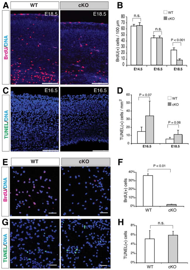
Figure 3.
In vivo and in vitro changes of proliferation and apoptosis in the β-catenin-cKO neocortex or dissociated neurosphere cells. (A, B): Significantly reduced BrdU(+) cells (per 100-μm-wide neocortical column) in E18.5 mutants. (C, D): Increased TUNEL(+) cells (per square mm neocortex) in E16.5 and E18.5 mutants. (E–H): Significantly reduced BrdU(+) percentage and no difference of TUNEL(+) percentage in the dissociated mutant sphere cells compared with those in the wild-type. n.s., no statistical difference (p > .05). Scale bars in A and C, 100 μm; scale bars in E and G, 50 μm.

