
Fig. 2. Amino acid sequences of the CDR2β regions of mouse Vβs illustrate residues shared between different Vβs.
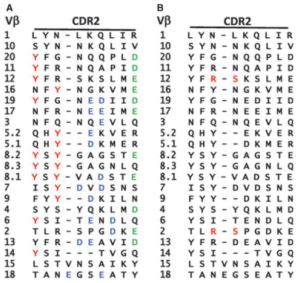
The amino acid sequences of the CDR2 regions of mouse Vβs are shown. (A) The Y46 Y48 tyrosine motif shared by relatives of the mouse Vβ8 family are highlighted in red. The acidic amino acids that often form salt bridges with basic amino acids in MHCII are colored green (D or E at position 54) or blue (other D or E in this region). (B) The R S motif that makes contact with MHC in two structures containing Vβ2 is colored red (39).