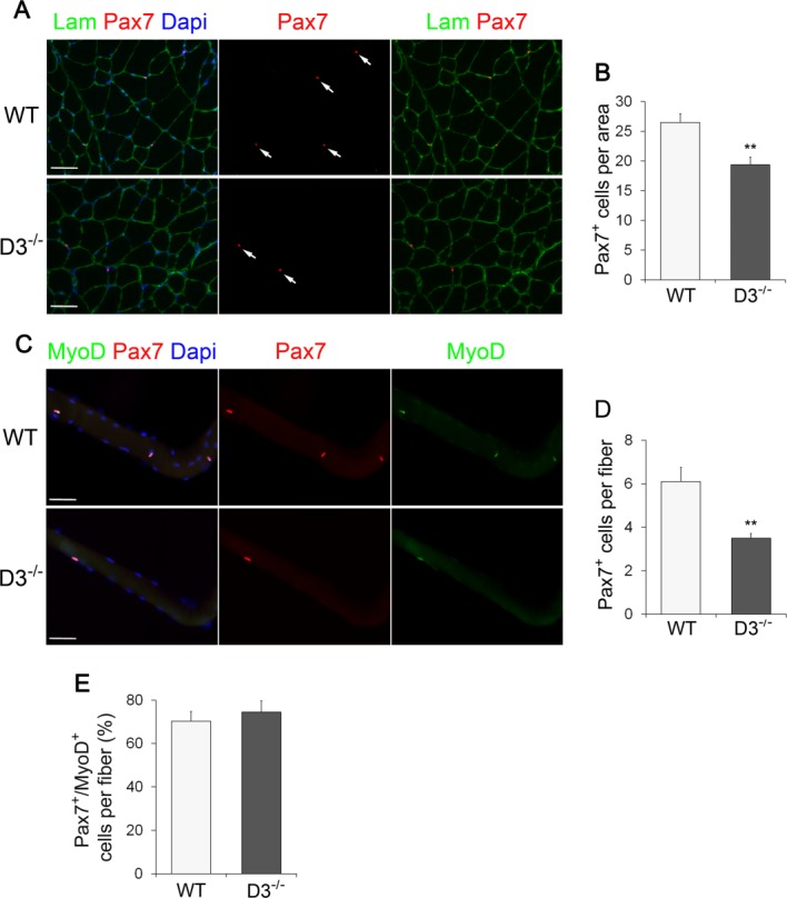
Figure 4.

Ablation of cyclin D3 leads to a decline in the adult satellite cell pool size. (A): Transverse sections of tibialis anterior muscles from WT and D3−/− male mice at P60 were stained with Laminin to identify myofibers (green) and Pax7 to identify sublaminar satellite cells (red) indicated by arrows. Nuclei were counterstained with Dapi (blue). (B): Average number of Pax7-positive cells per cross-sectional tissue area (mm2) in WT and D3−/− mice. Error bars represent ±SEM for n = 3. (C): Single myofibers freshly isolated from extensor digitorum longus muscles of 8–12-week-old WT and D3−/− mice were stained 6 hours ex vivo for Pax7 (red) and MyoD (green), and counterstained with Dapi (blue). (D): Average number of Pax7+ satellite cells per myofiber in WT and D3−/− mice. Error bars represent ±SEM for n = 5 WT and n = 7 D3−/−. (E): Quantification of Pax7+/MyoD+ satellite cells per myofiber in WT and D3−/− mice. Values are expressed as the mean percentage of Pax7+ cells that were MyoD-positive (means ± SEM for n = 4). Asterisks denote significance (**, p < .01). Scale bar = 50 µm. Abbreviations: Lam, Laminin; Dapi, 4′,6-diamidino-2-phenylindole; WT, wild type.
