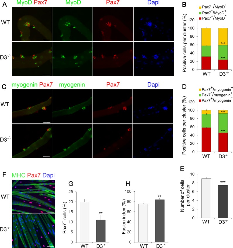
Figure 5.

Cyclin D3 loss affects SC differentiation and self-renewal. (A): Batches of single extensor digitorum longus (EDL) myofibers from WT and D3−/− mice were coimmunostained for Pax7 (red) and MyoD (green) after 72 hours in suspension culture. Counterstaining with Dapi (blue) was used to identify all nuclei present on the myofiber. (B): The graph represents the quantification of the Pax7+/MyoD−, Pax7+/MyoD+, or Pax7−/MyoD+ cells contained in clusters of four or more cells. Data are expressed as the percentage in each category of the total positive cells per cluster (means ± SEM). Clusters (270) from WT (n = 4) and D3−/− (n = 3) mice were analyzed, for a total of 2,000 cells counted for each genotype. (C): Myogenin (green) and Pax7 (red) coimmunostaining of WT and D3−/− EDL myofibers 72 hours after isolation, and counterstaining with Dapi (blue). (D): The graph represents the quantification of the Pax7+/Myogenin−, Pax7+/Myogenin+, or Pax7−/Myogenin+ cells. Data are expressed as in (B). Clusters (180) from WT (n = 3) and D3−/− (n = 5) mice were analyzed, for a total of 1,300 cells counted for each genotype. (E): Quantification of cluster size. The total number of cells present in the WT and D3−/− clusters analyzed in (B) was calculated and expressed as mean number of cells per cluster ± SEM. (F): Satellite cells derived from WT and D3−/− EDL myofibers were seeded in eight-well permanox chamber slides (1.2 × 10⁴/well) and transferred to DM after 24 hours. Cells were cultured in differentiation medium for 72 hours before fixing and immunostaining for MHC (green) to visualize myotubes and Pax7 to identify mononuclear undifferentiated reserve cells (red). Counterstaining with Dapi was used to visualize all nuclei (blue). (G): Relative number of Pax7-expressing cells. Values are expressed as the mean percentage ± SEM of total nuclei (WT: n = 6; D3−/−: n = 5). (H): Quantification of fusion index. Values are expressed as the mean ratio ± SEM of nuclei present in myotubes to the total number of nuclei (n = 5). Asterisks denote significance (**, p < .01; ***, p < .001). Scale bar = 50 µm. Abbreviations: Dapi, 4′,6-diamidino-2-phenylindole; MHC, myosin heavy chain; WT, wild type.
