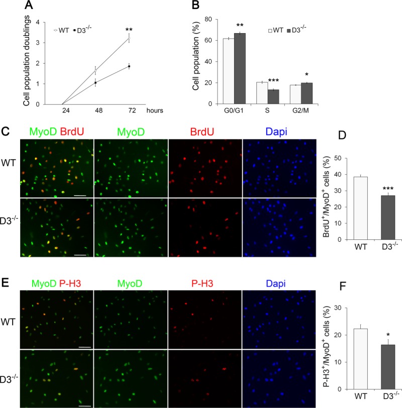
Figure 6.

Cyclin D3 null satellite cells show proliferative deficits in vitro. (A): Primary myoblasts from dissociated limb muscles of WT and D3−/− mice were seeded in eight-well permanox chamber slides (104/well) and counted in 10 independent microscopic fields after 24, 48, and 72 hours in culture (>95% myogenic cells, as assessed by MyoD staining). The graph represents the mean number ± SEM (n = 4) of cell population doublings at 48 and 72 hours, relative to 24 hours (log 2 [number of cells at 48 or 72 hours]/number of cells at 24 hours). (B): WT and D3−/− primary myoblasts cultured for 48 hours were detached and stained with propidium iodide. Flow cytometry analysis of the cell cycle reveals a lower percentage of D3−/− cells in S-phase and a higher percentage in G0/G1 and G2/M compared with WT cells (means ± SEM, n = 6). (C, E): WT and D3−/− primary myoblasts cultured for 48 hours were labeled with 10 µM BrdU for 4 hours before fixation and immunostaining to detect BrdU (red), MyoD (green), and PH3 (red) as a marker of mitotic cells. Nuclei were counterstained with Dapi (blue). (D, F): The percentages of BrdU or P-H3 positive cells/total MyoD-positive cells are shown. Error bars represent ±SEM (n = 4). Asterisks denote significance (*, p < .05; **, p < .01; ***, p < .001). Scale bar = 50 µm. Abbreviations: Dapi, 4′,6-diamidino-2-phenylindole; P-H3, Phospho-Histone H3; WT, wild type.
