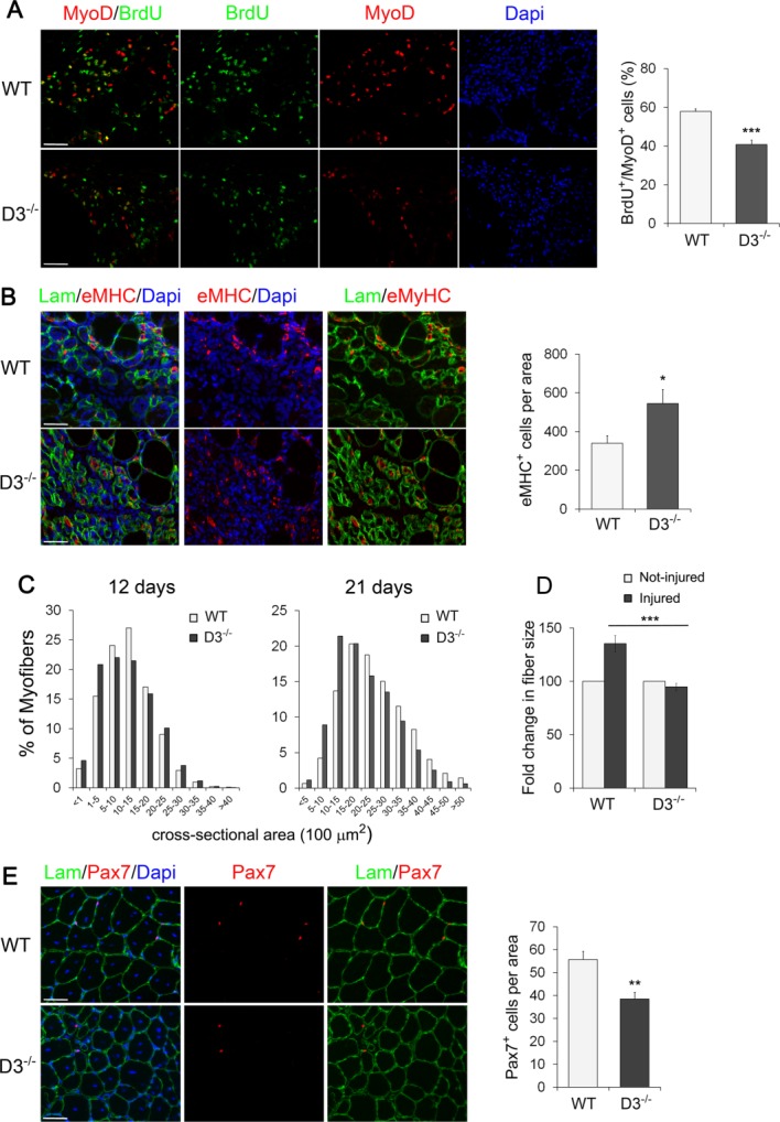
Figure 7.

Loss of cyclin D3 affects muscle regeneration. (A): Acute muscle injury was induced in WT and D3−/− male mice at P60 by intramuscular injection of cardiotoxin. Mice were injected BrdU intraperitoneally 6 hours before sacrifice. Left, cross-sections of tibialis anterior (TA) muscles after 3 days of regeneration stained for MyoD (red) and BrdU (green) to label proliferating myogenic progenitors. Nuclei were counterstained with Dapi (blue). The graph on the right represents the mean percentage ± SEM of MyoD-positive cells that were BrdU+ (WT: n = 5; D3−/−: n = 4). (B) Left, muscle cross-sections as in (A) were stained for eMHC (red) to identify newly generated myofibers and Laminin (green) to identify damaged fibers. Nuclei were counterstained with Dapi (blue). Right, quantitative analyses of nascent fiber number normalized to cross-sectional area of regenerating tissue (mean ± SEM for n = 4). (C) Frequency histograms showing the cross-sectional area of WT and cyclin D3-null myofibers 12 days and 21 days after cardiotoxin injection (12 days: n = 3; WT: 7,866 myofibers; D3−/−: 7,633 myofibers—21 days: n = 4; WT: 4,826 myofibers; D3−/−: 5,006 myofibers). (D): Fold change in regenerated fiber size from WT and D3−/− TA muscles 21 days after injury relative to uninjured fibers in adjacent areas of the tissue (means ± SEM, n = 4). (E): Left, cross-sections from regenerating TA muscles 21 days after injury stained for Pax7 (red) and Laminin (green). Nuclei were counterstained with Dapi (blue). Right, quantification of Pax7-positive cells associated with regenerated myofibers, normalized to 1 mm2 of tissue cross-sectional area (means ± SEM; WT, n = 5; D3−/−, n = 3). Asterisks denote significance (*, p < .05; **, p < .01; ***, p <.001). Scale bar = 50 µm. Abbreviations: Dapi, 4′,6-diamidino-2-phenylindole; eMHC, embryonic myosin heavy chain; Lam, Laminin; WT, wild type.
