Figure 4.
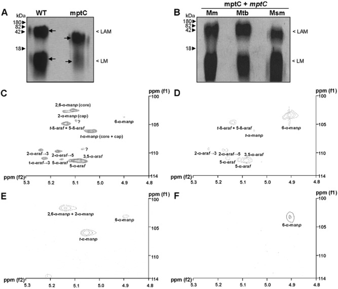
Analysis of lipoglycan profiles confirms disrupted mannan core branching in mptC mutant.A and B. Lipoglycans extracted from (A) wildtype M. marinum (WT) and the mptC mutant (mptC), or (B) the mptC mutant complemented with M. marinum, M. tuberculosis or M. smegmatis mptC (mptC + mptC Mm, Mtb and Msm respectively) were run on SDS-PAGE and visualized using Pro-Q emerald glycoprotein stain (Invitrogen). Arrows in A indicate ‘Centre of Maxima’ (CoM) of these heterogeneous lipoglycan species.C–F. Two dimensional 1H-13C-NMR heteronuclear multiple-quantum correlation (HMQC) spectra from lipoglycans from wildtype M. marinum and mptC mutant. LAM (C and D) and LM (E and F) were extracted and purified from M. marinum and the mptC mutant respectively, and their 2D 1H-13C-NMR HMQC spectra were recorded. Various chemical resonances were annotated to their respective linkages based on previous work (Nigou et al., 1999; Kaur et al., 2008).

