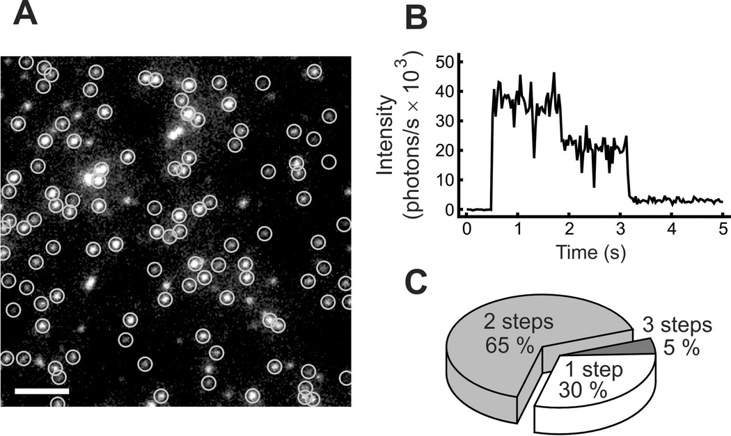
Figure 2.
Evidence that the human proton channel, hHV1, is a dimer. A shows GFP (green fluorescent protein) tagged hHV1 channels visualized under fluorescence microscopy. Circled spots were followed over time, as illustrated in B, where the fluorescence intensity of one spot can be seen to decay in two distinct steps. The pie chart in C shows the frequency that tagged channels decayed with the indicated number of steps. Reprinted from: Tombola F, Ulbrich MH, and Isacoff EY. The voltage-gated proton channel Hv1 has two pores, each controlled by one voltage sensor. Neuron 58, 546–556, copyright 2008, with permission from Elsevier.

