Figure 2.
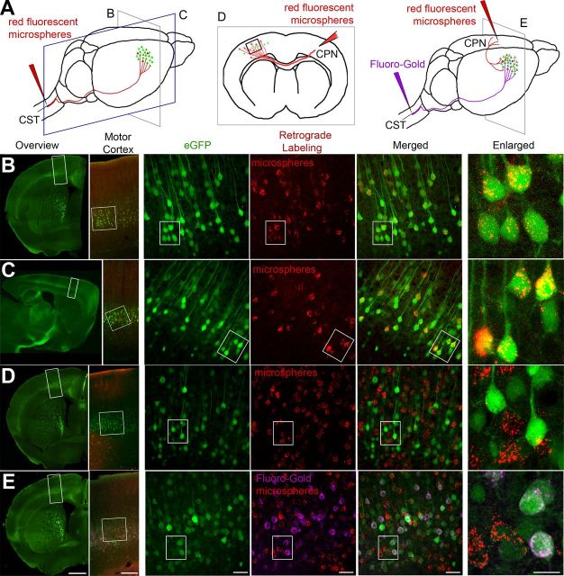
Retrograde labeling studies suggest CSMN identity for eGFP+ neurons in motor cortex. A, Drawing of experimental design. CSMN were retrogradely labeled by red fluorescent microsphere injection into the CST (red arrow, left). CPN were retrogradely labeled by red fluorescent microsphere injection into the contralateral hemisphere (red arrow, middle). CSMN (FG; purple arrow) and CPN (red fluorescent microspheres; red arrow, right) were labeled with two retrograde labeling surgeries. All mice were analyzed at P30. Coronal (B) and sagittal (C) sections are imaged to show colocalization of eGFP+ neurons with red fluorescent microspheres after retrograde labeling from CST. Boxed areas are enlarged to the right. D, eGFP+ neurons located in layer 5 of motor cortex do not contain red fluorescent microspheres after retrograde CPN labeling from the contralateral hemisphere. Boxed areas are enlarged to the right. E, eGFP+ neurons located in layer 5 of motor cortex contain FG, but not red fluorescent microspheres after dual retrograde labeling of CSMN (FG) and CPN (red fluorescent microspheres). Boxed areas are enlarged to the right. Images represent intrinsic eGFP expression without immunocytochemical enhancement. Scale bars (left to right), 1 mm, 250 μm, 50 μm, 50 μm, 50 μm, 20 μm.

