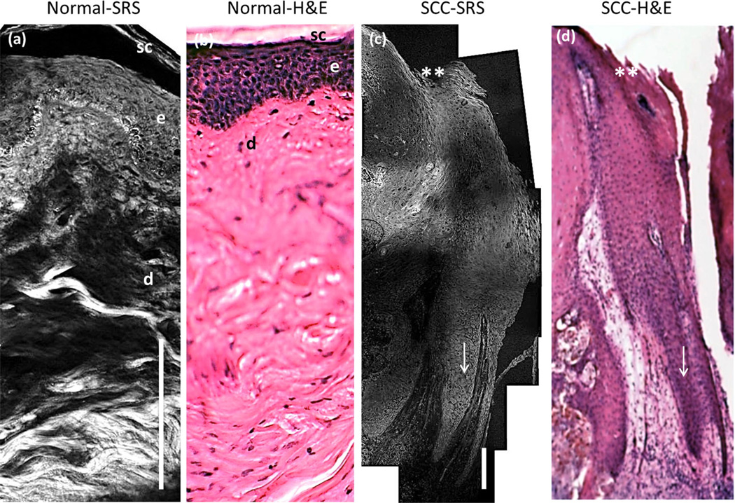
Fig. 3.
Comparison of SRS mosaic images of healthy human skin and superficial SCC ex vivo. a: SRS unstained tissue image of healthy human skin. sc, stratum corneum; e, epidermis; d, dermis. b: H&E stained specimen of an adjacent healthy skin section. c: SRS unstained tissue image of superficial SCC. d: H&E stained specimen of an adjacent SCC section. ** marks the skin surface; arrow indicates the keratinocytes pushing into the dermis. The imaging anatomical depth of healthy and SCC sample is 300 µm and 1.1 mm, respectively. Scale bar is 100 µm. Imaging performed using a 20X, 0.75NA Olympus objective lens.

