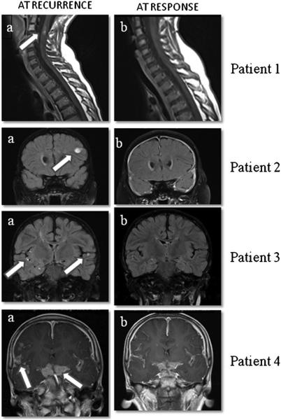
Fig. 1.
MRI images from four patients at the time of medulloblastoma recurrence and at response. Sagittal T1 post-contrast images of the cervicothoracic spine of patient 1 (top row) demonstrate enhancing intramedullary drop metastases (arrow in a at the level of C2–3, that resolve (b) after treatment. Coronal post-contrast T2 FLAIR images of patient 2 (second row) demonstrate an enhancing lesion in the left frontal lobe at recurrence (arrow in a) that resolves (b) after treatment. Coronal post-contrast T2 FLAIR images of patient 3 (third row) demonstrate leptomeningeal metastases in the bilateral sylvian fissures (arrows in a) and adjacent to the right hippocampus at recurrence that resolve (b) after treatment. Post-contrast T1 coronal images of patient 4 (fourth row) demonstrate thick leptomeningeal metastases in the suprasellar cistern and sylvian fissures (arrows in a) at the time of recurrence that significantly improve (b) following treatment

