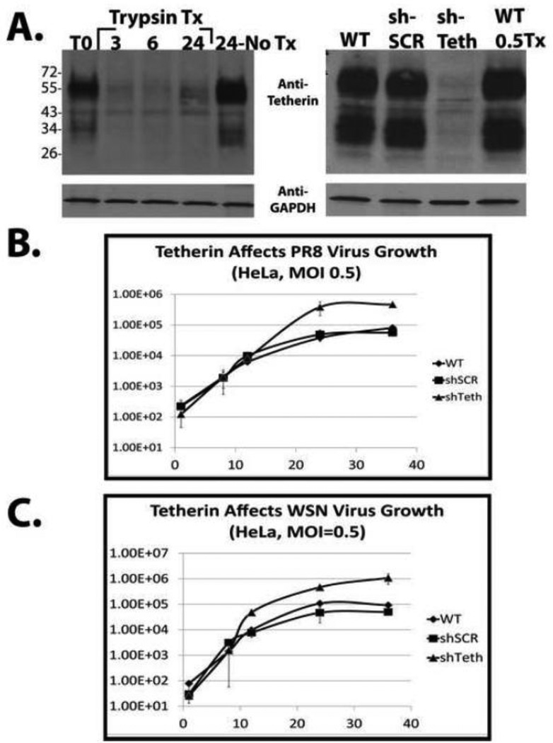
Figure 6. Tetherin contributes to the poor growth properties of influenza virus in HeLa cells.
Panel A, left, HeLa cells were washed 2× and the medium was either unchanged (control), or changed to standard influenza virus growth medium (trypsin-containing) as described in the text. Just prior to the addition of virus growth medium, T0, and at 3, 6, and 24 hours post medium change, cells were lysed and analyzed by western blot. Panel A, right, Cells were infected at moi = 0.05 and then incubated in the tetherin-sparing virus growth medium described in the results. 24 hours after medium transfer, the cells were lysed and the efficiency of shRNA-mediated stable tetherin knockdown was determined by western blot. Anti-Glyceraldehyde-3-phosphate dehydrogenase (GAPDH) antibody was used as a loading control. Panel B, Virus growth analysis performed after infection of the indicated HeLa cell lines with either WT PR8 (top) or WT WSN (bottom) viruses at an MOI of 0.5. Virus titration was performed by standard plaque assay on MDCK cells.

