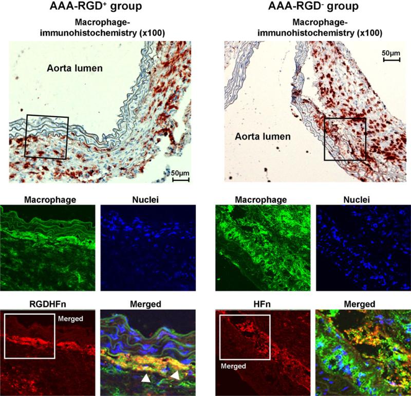
Fig. 5.
Macrophage immunostaining of representative abdominal aortic aneurysms (AAA). Immunohistochemical AAA staining showed mural macrophage infiltration, with immunofluorescence staining of the outlined region demonstrating the more intense RGD-HFn-Cy5.5 signal (red), colocalizing with medial and adventitial macrophages (green; arrowheads) as compared to HFn-Cy5.5 (see text for quantitative colocalization analysis).

