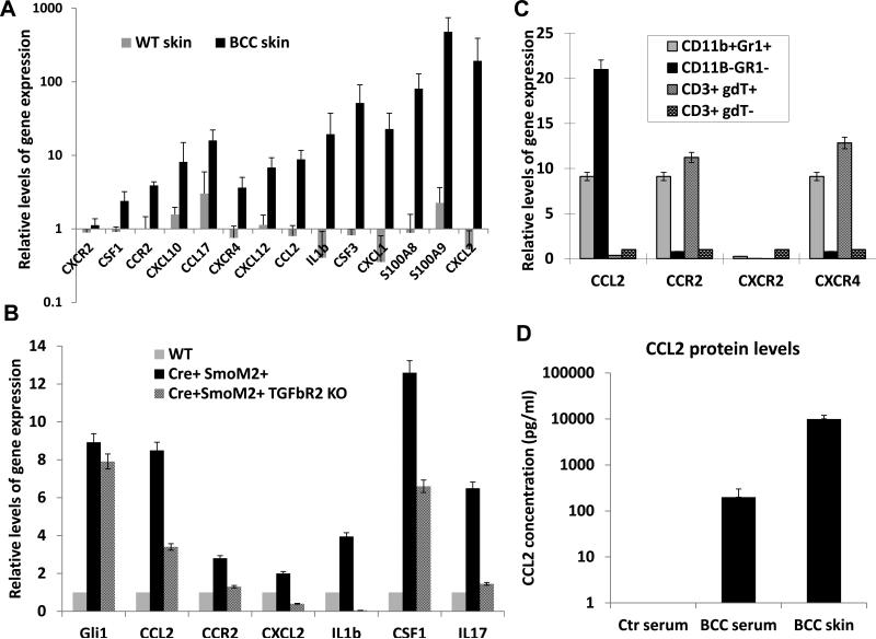
Figure 6. Expression of chemokines, cytokines and inflammatory factors in SmoM2-mediated skin tumors.
A shows altered expression of chemokines, cytokines and inflammatory factors in SmoM2-driven skin tumors. Comparison between K14-creER/R26-SmoM2 (as WT skin) mice and R26-SmoM2 mice (as BCC skin) showed significant elevation of gene expression (indicated by*) for Csf1, Csf3, Ccr2, Ccl2, cxcl10, Ccl17, Cxcr4, Cxcl12, Cxcl1, Cxcl2, Il1b, S100a8 and S100a9, but not Cxcr2. B shows examples of factors regulated by TGFβ signaling in skin tissues. Significant difference between normal and tumorous skin tissues was indicated as * whereas significant reduction between Mx1-cre/R26-SmoM2 and Mx1-cre/R26-SmoM2/Tgfbr2f/f mice was indicated as ** (p values< 0.05 by Student's t test). C shows expression of chemokines and their receptors in different cell populations of SmoM2-driven skin tumors. Significant elevation of gene expression was indicated by * (p values<0.05). D shows ELISA analysis of the CCL2 protein level in peripheral blood and skin tissues from tumor-bearing mice, with a comparison with the serum level of CCL2 from R26-SmoM2 mice (no tumor-bearing control). While control mice had nearly undetectable CCL2 protein in the serum, the serum level of CCL2 protein in tumor-bearing mice reached ~200pn/ml. The highest level was detected in the skin tumor (~1,000pg/ml). It thus appears that there is a CCL2 protein gradient from a low level in the serum to a high level in the tumor.

