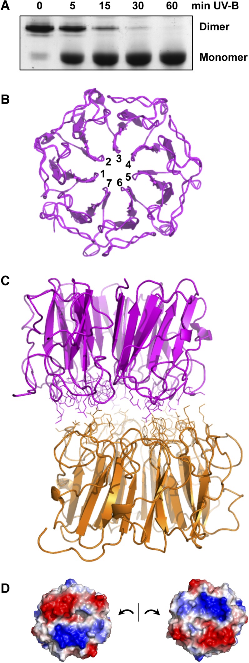
Figure 1.
UVR8 Dimer Structure and Monomerization.
(A) UV-B induces monomerization of UVR8. Coomassie blue–stained SDS-PAGE gel of purified UVR8 exposed for the times shown to 1.5 μmol m−2 s−1 narrowband UV-B (λmax 311 nm). Samples were prepared for electrophoresis without boiling. The UVR8 dimer and monomer are indicated.
(B) Seven-bladed β-propeller structure of the UVR8 monomer. The structure is shown for amino acids 14 to 380.
(C) Structure of the UVR8 dimer showing residues at the dimer interaction surface.
(D) The dimer interaction surfaces of two UVR8 monomers displayed to show patches of complementary electrostatic potential. Basic (blue) and acidic (red) amino acids contribute positive and negative charges, respectively.
Images in (B) to (D) were produced using PyMOL. (All panels produced from data presented by Christie et al. [2012].)

