Figure 3.
UVR8 Signaling and Regulation.
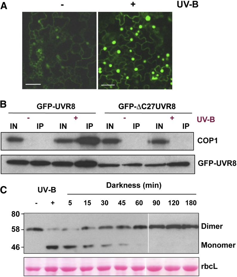
(A) UV-B–induced nuclear accumulation of UVR8. Confocal image of epidermal cells of Arabidopsis uvr8-1 plants expressing UVR8pro:GFP-UVR8. Plants were grown in 20 μmol m−2 s−1 white light lacking UV-B (−) and exposed to 3 μmol m−2 s−1 broadband UV-B for 4 h (+). Bar = 20 μm. (Modified from Kaiserli and Jenkins [2007], Figure 3A.)
(B) Interaction of UVR8 and COP1 in plants. Whole-cell protein extracts were obtained from uvr8-1/UVR8pro:GFP-UVR8 and uvr8-1/UVR8pro:GFP-ΔC27UVR8 plants treated (+) or not (−) with 3 μmol m−2 s−1 narrowband UV-B. Coimmunoprecipitation assays were performed under the same illumination conditions. Input samples (15 μg; IN) and eluates (IP) were fractionated by SDS-PAGE, and protein gel blots were probed with anti-COP1 and anti-GFP antibodies. (Reprinted from Cloix et al. [2012], Figure 3A.)
(C) Regeneration of the UVR8 dimer. Immunoblot of whole-cell protein extracts from wild-type Landsberg erecta plants probed with anti-UVR8 antibody. Plants were exposed to 2.5 μmol m−2 s−1 narrowband UV-B for 3 h (+ UV-B) and then transferred to darkness for the indicated time periods before extracts were made. Extract samples were prepared for electrophoresis without boiling prior to SDS-PAGE and immunoblotting. The UVR8 dimer and monomer are indicated. Ponceau staining of Rubisco large subunit (rbcL) is shown as a loading control. (Reprinted from Heilmann and Jenkins [2013], Figure 1A.)

