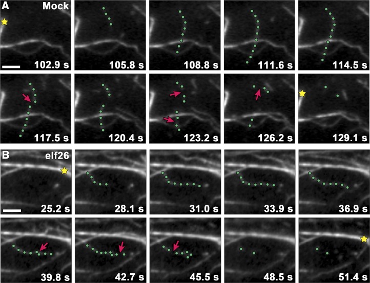
Figure 3.
Filament Severing Is Significantly Reduced following elf26 Treatment.
(A) Time-lapse series of VAEM images shows actin filament turnover in the cortical cytoplasm of a mock-treated epidermal cell. The highlighted filament (green dots) elongated at 1.72 µm s−1 before suffering several breaks (arrows). Actin filament bundles (stars) remained relatively stationary throughout the time series.
(B) A representative growing filament (green dots) from a wild-type cell treated with 1 μM elf26 peptide for 5 min displayed fewer severing events (arrows) compared with the mock-treated cell.
Micrographs in (A) and (B) were collected at 1.5-s intervals, and every other image is presented in each montage. Bars = 5 μm.

