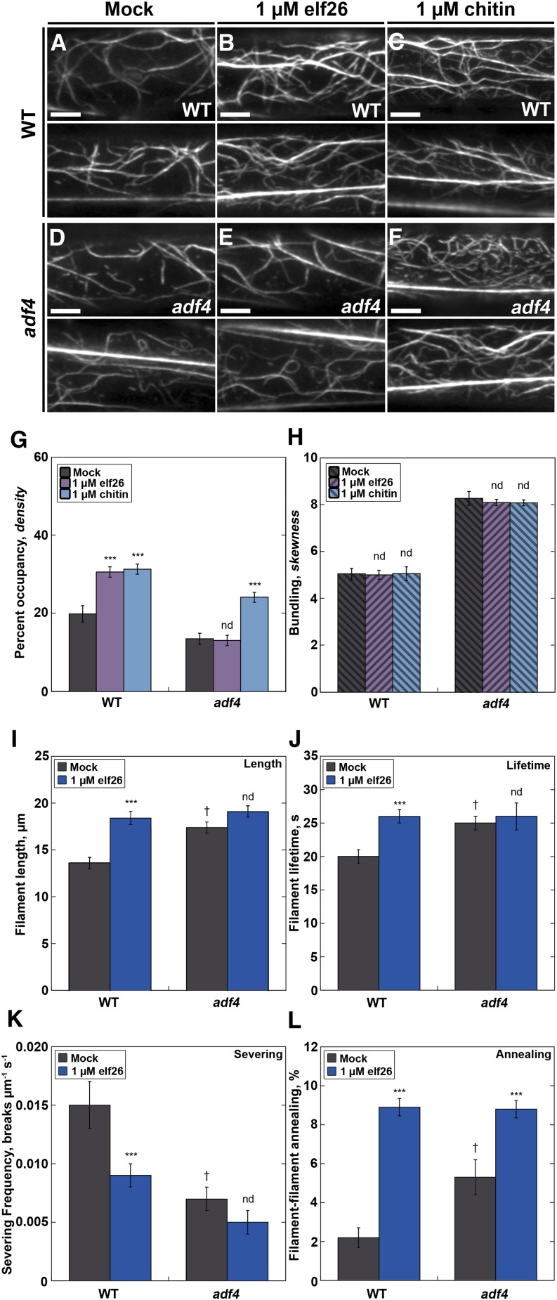
Figure 4.
The adf4 Mutant Lacks Changes in Actin Filament Architecture and Dynamics following Treatment with elf26.
(A) to (F) Actin filaments in mock-treated epidermal cells from the adf4 mutant (D) appeared to be significantly less abundant and more bundled compared with wild-type cells. Notably, the actin architecture in adf4 cells did not seem to change following treatment with elf26 peptide (E), unlike wild-type cells (B), where actin filaments appeared to be more abundant after MAMP treatment. In response to the fungal MAMP, chitin, cortical actin abundance increased in both the wild type (C) and the adf4 mutant (F). Bars = 5 µm.
(G) Actin filament abundance was measured in epidermal cells at the base of wild-type and adf4 mutant hypocotyls after treatment with MAMPs for 5 min. Wild-type epidermal cells treated with 1 µM elf26 or 1 µM chitin had a significant increase in filament abundance compared with mock-treated seedlings. Actin filament abundance in cells from the adf4 mutant treated with 1 µM elf26 remained unchanged compared with mock-treated controls. However, actin filament abundance in the adf4 mutant was significantly elevated following treatment with chitin.
(H) The extent of actin filament bundling was not altered following treatment with chitin in either wild-type or adf4 hypocotyl epidermal cells. The same images analyzed in (G) were measured for actin filament bundling.
(I) to (K) Several parameters of actin filament turnover do not change in the adf4 mutant following elf26 treatment. The adf4 mutant had significantly enhanced filament lengths (I) and lifetimes (J) as well as a reduction in severing frequency (K) compared with the wild type. Whereas each of these parameters was significantly changed in wild-type seedlings upon treatment with elf26, these do not change in the adf4 mutant.
(L) By contrast, there were significant increases in filament-filament annealing in the adf4 loss-of-function mutant compared with wild-type cells. Following elf26 treatment, both the wild type and the adf4 mutant respond with significantly enhanced filament–filament annealing.
Values given are means ±se (n = 300 cells per treatment and genotype from at least 30 hypocotyls). Asterisks represent significant differences by ANOVA, with Tukey HSD posthoc analysis (nd = not significantly different from genotype-specific mock control; ***P < 0.001; † denotes significant differences between the wild type and adf4). For more details, see Supplemental Table 1.

