Figure 2.
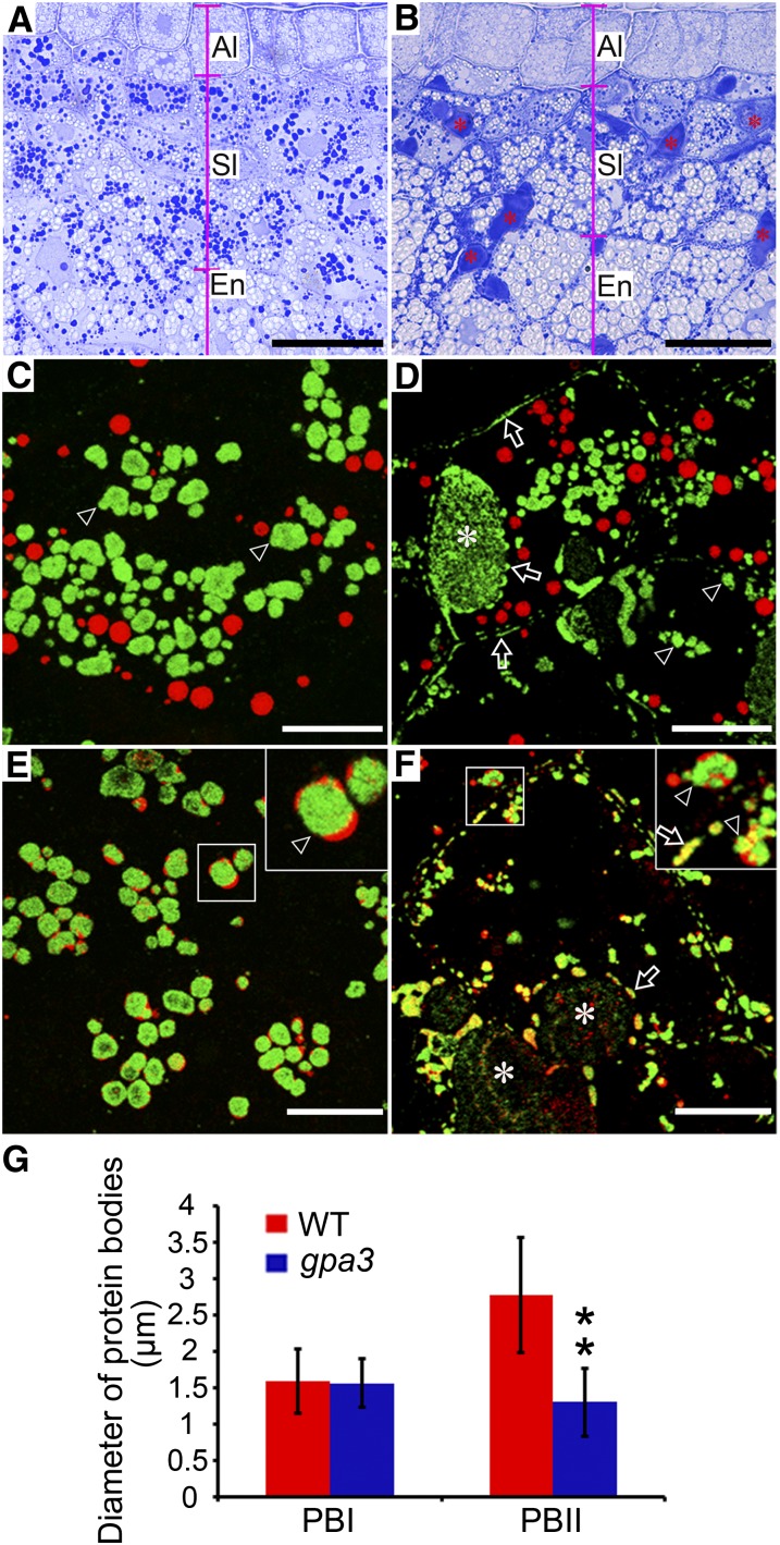
Light and Immunofluorescence Microscopy Images of Protein Bodies in the Subaleurone Cells of the Wild Type and the gpa3 Mutant.
(A) and (B) Light microscopy observation of wild-type (A) and gpa3 mutant (B) grain sections stained with Coomassie blue. Note the numerous PMB structures (asterisks) present in the gpa3 mutant. Al, aleurone layers; Sl, subaleurone layers; En, starchy endosperm. Bars = 25 μm.
(C) to (F) Immunofluorescence microscopy images of storage proteins in wild-type ([C] and [E]) and gpa3 mutant ([D] and [F]) grains. Secondary antibodies conjugated with Alexa fluor 488 (green) and Alexa fluor 555 (red) were used to trace the antigens recognized by the antiglutelin and antiprolamin antibodies, respectively, in (C) and (D). Similar reactions were performed with anti-α-globulin antibodies instead of antiprolamin antibodies in (E) and (F). The insets represent magnified images of the selected areas in (E) and (F). Arrows indicate the protein granules lying along the cell periphery and PMB structures (asterisks), while arrowheads indicate the PBIIs. Bars = 10 μm.
(G) Measurement of the diameters of PBIs and PBIIs. Values are means ± sd. **P < 0.01 (n > 300, Student’s t test).

