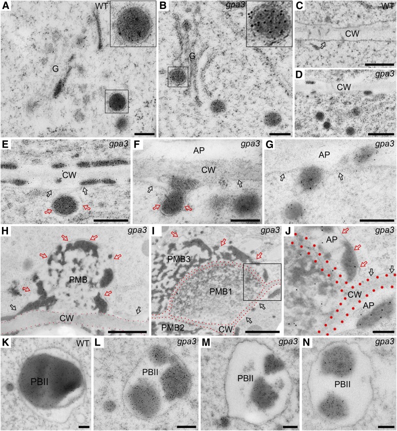
Figure 3.
Immunoelectron Microscopy Localization of Glutelins Depicting the Biogenesis of PMBs in the gpa3 Mutant.
Ultrathin sections of developing wild-type and gpa3 subaleurone cells were prepared using an HPF procedure, and developing wild-type and gpa3 subaleurone cells were labeled with antiglutelin primary antibodies and gold colloid–conjugated secondary antibodies. Gold particles were observed in the Golgi (G), DVs, protein granules in the apoplast (AP), PMBs, and PBIIs. Black arrows indicate the PM, while red arrows indicate the envelope of the PMB. Red dots outline the cell wall (CW) region. Bars in (A) to (C), (E) to (G), and (J) to (N) = 200 nm; bars in (D) = 500 nm; bars in (H) and (I) = 1 mm.
(A) and (B) Electron micrographs showing that, as in the wild type (A), DVs can normally bud off from the Golgi in the gpa3 mutant (B). Note that DVs are enclosed with a single membrane (insets). The insets represent the magnified images of the selected DVs.
(C) and (D) General overview of cell periphery in developing wild-type (C) and gpa3 (D) subaleurone cells. Note that numerous glutelin-containing DVs accumulate near the PM.
(E) to (G) Electron micrographs showing that single membrane (red arrows)–enclosed DVs can fuse with the PM (black arrows) and expel their contents into the apoplast.
(H) to (J) Electron micrographs showing young (H) and mature (I) PMB structures. Note that the young PMB has a single membrane envelope (red arrows), which is physically connected with the PM (black arrows). (J) represents the magnified image of the selected area in (I).
(K) to (N) Electron micrographs showing that DVs can fuse with PSVs to form the PBIIs in the wild type (K) and the gpa3 mutant ([L] to [N]), but the PBII appears smaller in the gpa3 mutant.

