Figure 5.
Subcellular Localization of GPA3 in Arabidopsis Protoplasts and Developing Subaleurone Cells.
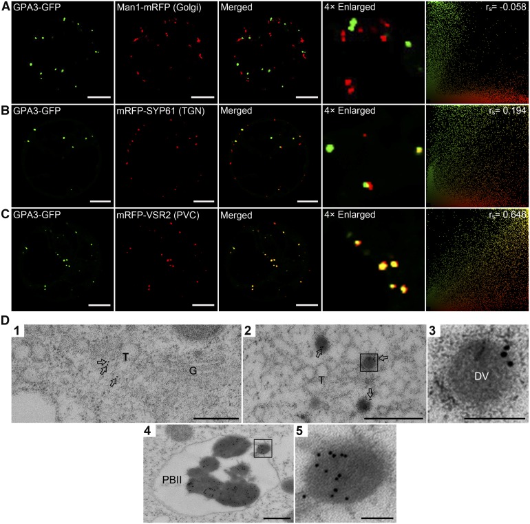
(A) to (C) Confocal microscopy images showing that GPA3-GFP is localized as punctate signals in the cytosol and its distribution is obviously distinct from the marker for Golgi (Man1-mRFP [A]) but partially overlaps with the markers for TGN (mRFP-SYP61 [B]) and PVC (mRFP-VSR2 [C]). PSC coefficients (rs) between GPA3-GFP and each marker are shown in the right panels. Bars = 10 μm.
(D) Immunoelectron microscopy localization of GPA3 in developing subaleurone cells. Ultrathin sections prepared using HPF subaleurone cells of transgenic plants expressing P35S:GPA3-GFP were labeled with anti-GFP antibodies, where gold particles (arrows) are found in the TGN (T; panel 1), DVs (panels 2 and 3), and PBIIs (panels 4 and 5). Panels 3 and 5 are the magnified images of selected areas in panels 2 and 4, respectively. G, Golgi. Bars in panels 1, 2, and 4 = 500 nm; bars in panels 3 and 5 = 100 nm.

