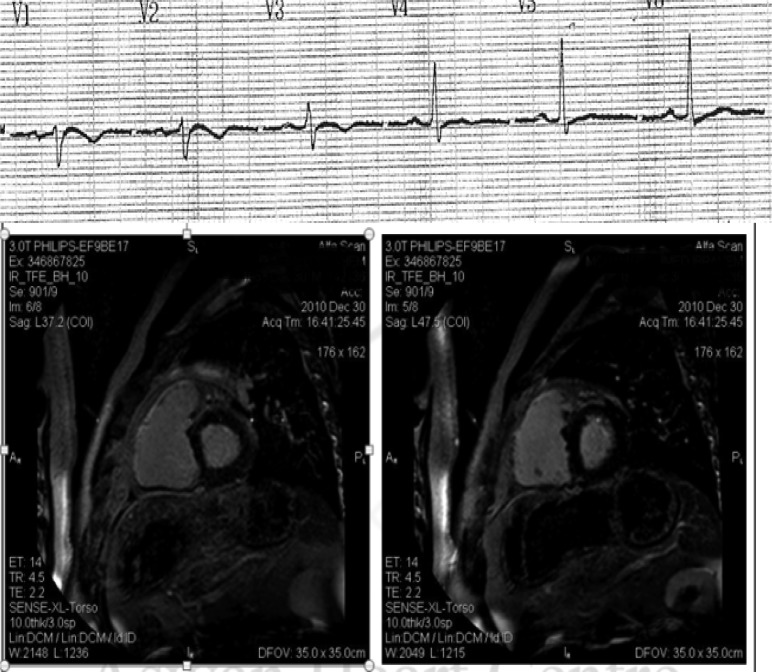
Figure 6. .
Above: Surface ECG precordial lead recordings of a 50 year old Egyptian/Sudanese patient showing Epsilon wave in V1, T wave inversion in V1-3, and relative QRS prolongation in right precordial leads. Below: Cardiac magnetic resonance imaging demonstrating delayed gadolinium enhancement in the right ventricular free wall denoting extensive fibrosis. The patient suffered two episodes of sustained ventricular tachycardias with left bundle branch block pattern with altered level of consciousness. An ICD was implanted for secondary prevention. (Images courtesy of Mohamed Donya, MD, FRCR, Aswan Heart Centre, Aswan, Egypt).

