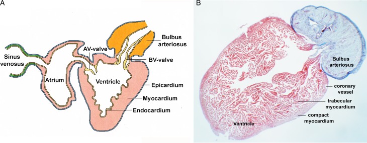
Figure 1. .
Anatomy of the adult zebrafish heart. (A) Schematic representation of the adult zebrafish heart. (B) Trichrome stained histological section through the ventricle and bulbus arteriosus, which labels myocardium in red whereas nonmuscle tissue and extracellular matrix in blue. Note the trabecular layer, which is extensive in the adult zebrafish heart, while the compact layer makes only a minor fractor of the total mass of the ventricular wall. Rarely coronary vessels are observed in the compact layer mocardium.

