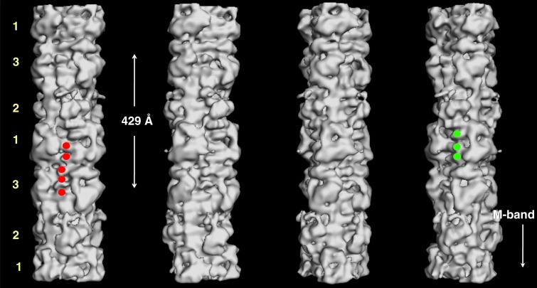
Figure 15. .
3D reconstruction of myosin filament from normal un-diseased human heart muscle under relaxing conditions (from 24), rotated by 30° around the filament long axis so that to show the different features on the surface of the filament corresponding to the myosin heads, titin and MyBP-C. Three C-terminal domains of C-protein (domains C8 to C10) next to myosin heads of level 1 (labelled by green circles in the fourth panel). To the left side of C-protein domain, another set of domain densities are observed which represent titin (five of which are labelled by red circles in the first panel). The crown level numbers are labelled. The direction of the barezone region is towards the bottom.

