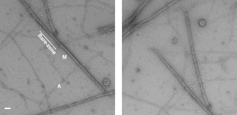
Figure 8. .
Electron microscopy images of myosin filaments isolated from human heart muscles in the normal undiseased state and under relaxing conditions (24) taken at a magnification of 29,000x. This shows intact myosin filaments (M) with identified barezone regions with their centres. It also shows actin filaments (A) in the background. Scale bar is 2000 Å.

